BCT Press Release
Skip Ribbon Commands
Skip to main content
Sign In
{{'GLOBAL_MESSAGE_HEADLINE' | translate}}
{{'GLOBAL_MESSAGE_CHOOSE_LANG' | translate}}
en
日本語
中文
{{'GLOBAL_MESSAGE_OR' | translate}}
{{'GLOBAL_MESSAGE_SET_COUNTRY' | translate}}
{{'ELOQUA_BANNER_DECLINE_CONFIRM' | translate}}
{{'ELOQUA_BANNER_DECLINE_CONFIRM_CLOSE' | translate}}
{{'ELOQUA_BANNER_ACCEPT' | translate}}
{{'ELOQUA_BANNER_DECLINE' | translate}}

Spot it: How to identify clumping in collection procedures

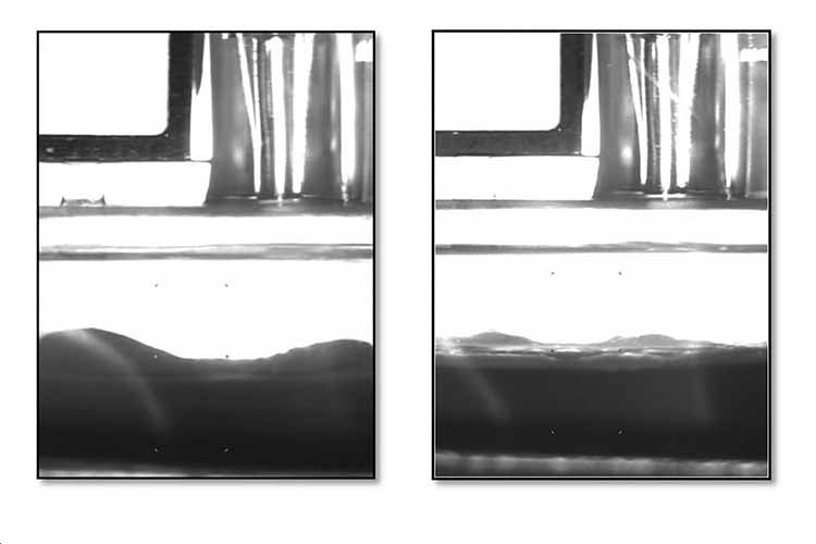
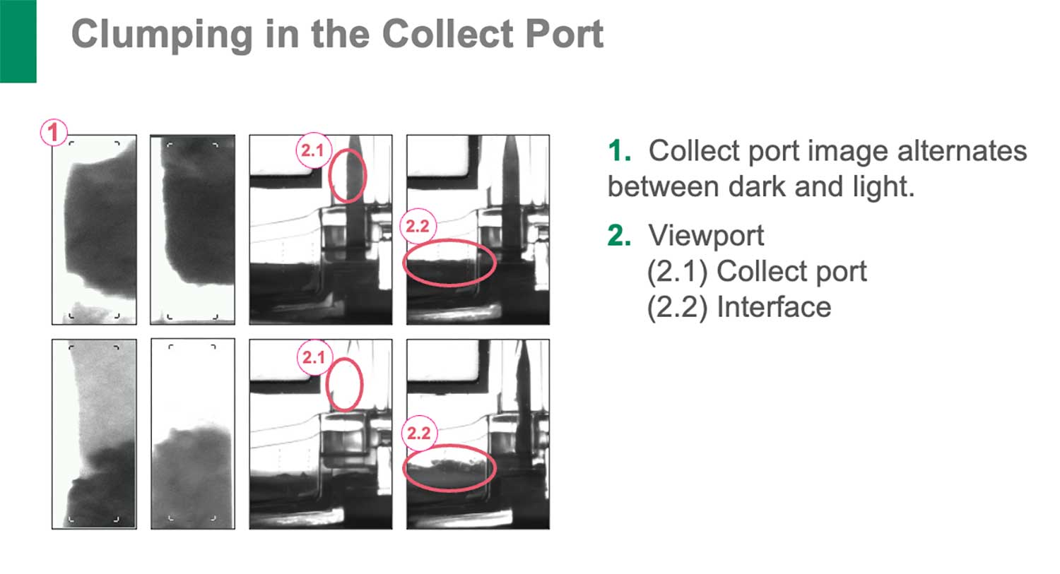
​​​When clumping occurs during a collection procedure on Spectra Optia, it can look like there are hills or peaks at the connector interface, instead of a straight, defined separation of cellular layers. You might see platelet aggregates exiting the centrifuge through the collect line. The color in the collect line may be unstable and go from light to dark and back again, without any changes being made to the collection preference.


The photo below shows what clumping can look like in a collection procedure when you are looking through the viewport at the connector.

The photos below are examples of what you might see when looking at the AIM system if there is clumping in the collect port.


To learn more best practices for managing and resolving clumping in collection procedures, check out these additional resources:


Source:
Terumo Blood and Cell Technologies. Continuous Mononuclear Cell Collection (Version 12 CMNC) Procedure Training. 2023. Part number TS-OPTI- 00124.

{{'SEARCH_MODAL_TITLE' | translate }}
{{'SEARCH_MODAL_OR' | translate }}

{{'SEARCH_MODAL_BROWSE' | translate }} {{'SEARCH_MODAL_ENLABEL' | translate }}
{{'SEARCH_MODAL_CHOOSE_LANGUAGE' | translate }} {{'SEARCH_MODAL_CHANGE_LANGUAGE' | translate }}
{{$index+1}}. {{s.label}}